Znieczulenie komputerowe transkortykalne w zarządzaniu opieką w nagłych wypadkach

W nagłych wypadkach stomatologicznych, zastosowanie znieczulenia miejscowego do leczenia bólu o pochodzeniu endodontycznym wykazuje coraz wyższy odsetek niepowodzeń. Zespół Oddziału Stomatologii Odtwórczej i Endodontyki Szpitala Uniwersyteckiego w Marsylii zakończył prospektywne badania kliniczne w celu oceny skuteczności znieczulenia transkortykalnego za pomocą systemu QuickSleeper® po niepowodzeniu w zastosowaniu znieczulenia klasycznego.

Leczenie bólu w zarządzaniu nagłymi wypadkami stomatologicznymi jest wyzwaniem dla klinicysty, szczególnie, gdy ból ten jest związany z patologią endodontyczną. W rzeczywistości, jeśli znieczulenie miejscowe (blokada nerwu zębodołowego dolnego przy otworze żuchwowym) daje bardzo dobre wyniki w odniesieniu do planowanej opieki dentystycznej na zębie bezobjawowym, ze wskaźnikiem skuteczności wahającym się od 75 do 90% (1), wskaźnik niepowodzenia tych samych środków znieczulających istotnie wzrasta w przypadku nieodwracalnego zapalenia miazgi lub tkanek okołowierzchołkowych. Stąd też, badania kliniczne wykazały, że znieczulenie w postaci samej blokady IAN było nieskuteczne w 30 do 80% przypadków po zdiagnozowaniu zapaleniu miazgi na zębach trzonowych żuchwy (2, 3, 4).

Sformułowano kilka założeń mających na celu wyjaśnienie mechanizmów tych niepowodzeń (5), między innymi:

  • zmiany anatomiczne i kompleksowość unerwienia zęba;
  • zjawisko tachyfilaksji (redukcja odpowiedzi na substancję farmakologiczną związana z poprzednią iniekcją tej samej substancji);
  • wpływ zapalenia na pH otoczenia; kwasica zapalna w tkankach utrzymuje wysoki odsetek cząsteczek znieczulenia w formie jonizowanej, co sprawia, że nie są one w stanie przejść przez błonę plazmatyczną komórek a więc, nie są w stanie zablokować kanały sodowe;
  • wpływ zapalenia na krążenie krwi: wazodylatacja indukowana przez przekaźniki zapalenia prowadzi do spadku stężenia znieczulenia miejscowego;
  • wpływ zapalenia na reaktywność receptorów bodźców urazowych, prowadzący do ich nadmiernej pobudliwości;
  • czynniki psychologiczne (strach i niepokój powodują nadwrażliwość);
  • słabe opanowanie techniki znieczulania: jeśli wszystkie powyższe czynniki mogą wyjaśnić niepowodzenie, wydaje sie, że niska wiedza techniczna jest częstą przyczyną nieodpowiedniego znieczulenia po znieczuleniu blokadą IAN w zębach trzonowych żuchwy.

Stąd też, biorąc pod uwagę trudności kliniczne w zakresie uzyskania skutecznego znieczulenia w sytuacjach nagłych, parametr ten razem z brakiem doświadczenia studentów pracujących w szpitalach uniwersyteckich, celem tego badania prospektywnego była ocena skuteczności uzupełniającego znieczulenia doszpikowego z systemem QuickSleeper® po niepowodzeniu znieczulenia konwencjonalnego (IANB lub znieczulenie nadokostnowe) przeprowadzonego przez studenta. Oprócz jakości znieczulenia, oceniono igły oraz wkłady niezbędne dla każdego znieczulenia a także zadowolenie pacjenta.

Ta ocena kliniczna trwała sześć miesięcy i została przeprowadzona w “Unite Fonctionnelle Gaston Berger – AP – HM – Pole odontologique” w Marsylii.

Materiał i metoda

Grupa 25 pacjentów została zrekrutowana do tego badania. Wszyscy pacjenci przyszli i konsultowali się na Oddziale Ratunkowym.

Kryteria włączenia do badania: niepowodzenie przy pierwszym znieczuleniu przeprowadzonym przez studenta.

Kryteria wyłączenia: wstrzyknięcie sześciu wkładów, bez względu na technikę.

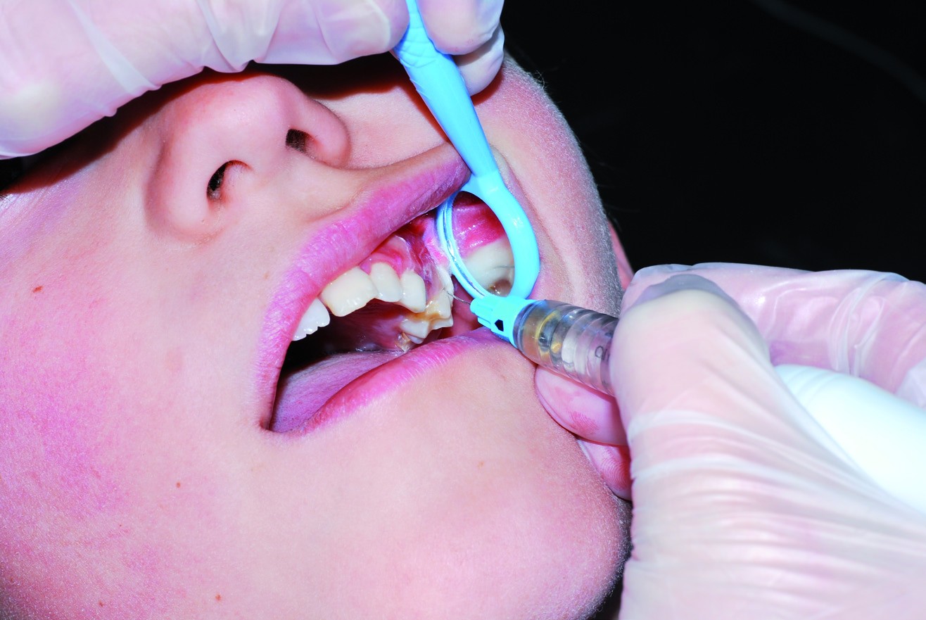
Po upewnieniu się o niepowodzeniu pierwszego znieczulenia w związku z brakiem możliwości przeprowadzenia zabiegu chirurgicznego w celu złagodzenia bólu pacjenta, przeprowadzone zostało uzupełniające znieczulenie transkortykalne przez instruktora z użyciem QuickSleeper® (formuła znieczuleniowa z 1: 100 000 epinefryny). Wyznaczono 23 parametry związane z realizacją znieczulenia transkortykalnego (liczba igieł, wkładów, złamania igieł, …) a także możliwości leczenia bezbólowego: wszystkie zostały zarejestrowane w formularzu klinicznym dla każdego pacjenta. Znieczulenie zostało uznane za skuteczne wtedy, gdy cały zabieg terapeutyczny mógł być zakończony a uznane za nieskuteczne, jeśli tak się nie stało.

Znieczulenie komputerowe transkortykalne: przypomnienie
W znieczuleniu transkortykalnym, iniekcja jest realizowana bezpośrednio do kości gąbczastej przez warstwę korową. W żuchwie, dyfuzja odbywa się głównie w kierunku środkowym od punktu iniekcji; zalecane jest wykonywanie iniekcji odśrodkowo względem leczonego zęba. Po iniekcji, roztwór ze znieczuleniem rozchodzi się w przestrzeniach szpikowych; liczba zębów, na które działa znieczulenie zależy od ilości wstrzykniętego roztworu. Znieczulenie doszpikowe odbywa się w trzech etapach. Znieczulenie śluzówki jest realizowane w zrośniętym dziąśle przy głębokości penetracji igły sięgającej zaledwie kilku dziesiętnym milimetra. Perforacja warstwy korowej jest całkowicie bezbolesna, ponieważ kość korowa nie jest unerwiona. Penetracja doszpikowa igły nie może nigdy przekroczyć 4 do 5mm w żuchwie. Wtedy możliwe jest powolna iniekcja roztworu.

Wyniki i dyskusja

W trakcie tej oceny, wskaźnik skuteczności uzupełniającego znieczulenia transkortykalnego wynosił 100%: dla każdego pacjenta, właściwy zabieg chirurgiczny mógł być zrealizowany bez potrzeby podawania dalszego znieczulenia.

Rozkład patologii i znieczuleń

Diagnoza Przekrwienie miazgi Zapalenie miazgi Inne
Redystrybucja 12 11 2
Znieczulenie transkortykalne 17
Znieczulenie PDL 8
Górne VS dolne zęby 11 VS 24

Niepowodzenia znieczuleń podstawowych dotyczyły prawie wyłącznie dolnych zębów trzonowych, jako, że w tym sektorze, grubość kości korowej sprawia, że znieczulenie nadokostnowe jest całkowicie nieskuteczne i wymaga iniekcji dalszej (blokada IAN), co może mieć niestałe wyniki, szczególnie w przypadku studenta, co zostało wcześniej zauważone.

Student wykonywał średnio iniekcje 3 do 4 wkładów i zużył 3 igły, ponieważ instruktor był często wzywany do pomocy po IANB, jednym znieczuleniu nadokostnowym i jednym znieczuleniu PDL. Odwrotnie, przy technice doszpikowej, wstrzyknięcie już 3/4 wkładu było średnio wystarczające do uzyskania głębokiego i natychmiastowego znieczulenia, które bezzwłocznie ulżyło pacjentowi.

Zdarzenia związane z zastosowaniem QuickSleeper®

Brak możliwości perforacji warstwy korowej Zmiana igły Złamanie igły Zatkanie się igły Znieczulenie tkanki miękkiej wargowej Czyszczenie igły
3 1 1 5 3 6

Zastosowanie QuickSleeper® okazało się być dość łatwe po okresie szkoleniowym, który był niezbędny do opanowania tej techniki.

System ten jest wyjątkowo wiarygodny, ponieważ jedynym zauważalnym przypadkiem było złamanie igły. W przypadku oddzielenia się igły, co miało miejsce przy rdzeniu igły, i wyciągnięciu odłączonego fragmentu jest to bardzo proste i nie powinno nastręczać obaw.

Z drugiej strony, należy zauważyć, że czasami perforacja korowa prowadzi do zatkania się igły, ponieważ ta sama igła jest używana do przekłuwania i iniekcji. Należy wtedy wyciągnąć igłę z kości korowej w celu oczyszczenia jej tak, aby została odetkana.

Pacjenci stwierdzili, że system ten jest bardzo wygodny i nie jest traumatyczny. Nie zgłoszono żadnych bolesnych przypadków pozabiegowych po zastosowaniu takiego znieczulenia doszpikowego. Tylko hałas silniczka czasami przestraszył pacjentów.

Wnioski

Przy uzupełniającym znieczuleniu transkortykalnym przeprowadzanym z użyciem systemu QuickSleeper®, wskaźnik skuteczności wynosił 100%. Umożliwiło to, we wszystkich przypadkach, wykonanie leczenia nagłego wymaganego w celu zapewnienia ulgi pacjentowi pomimo wyjątkowo niekorzystnego kontekstu związanego z bólem zęba a także stresem generowanym przez poprzednie nieudane znieczulenie. Wyniki te są zgodne z danymi pozyskanymi z literatury: znieczulenie doszpikowe, jako uzupełnienie do znieczulenia blokującego, zapewnia kompletne znieczulenie stomatologiczne w 100% przypadków na zębach bezobjawowych (6) oraz w 86 do 88% przypadków zębów z zapaleniem miazgi (88%). Liczba igieł i wkładów została podzielona przez cztery, tak więc znieczulenie transkortykalne wydaje się być odpowiednim do zastąpienia w sposób korzystny wielu powtarzanych iniekcji, które są klinicznie niepotrzebne i bolesne dla pacjenta.

Krzywa uczenia się jest istotna w celu opanowania zarówno urządzenia jak i techniki znieczulania transkortykalnego; brak doświadczenia może powodować pewną niewygodę dla operatora. Po zakończeniu tego okresu, używanie QuickSleeper® jest względnie łatwe. System znieczulenia doszpikowego taki, jak QuickSleeper® jest, więc wyjątkowo użyteczny w Oddziałach Ratowniczych Ośrodka Uniwersyteckiego.

Po tym pierwszym zachęcającym badaniu prospektywnym powinno nastąpić randomizowane, kontrolowane badanie mające na celu walidację tych wyników wstępnych. Ewolucja znieczulenia do śródkościa, znieczulenia typu osteocentral, będą, więc celem tych nowych badań.

Autorzy:

  • Dr Guillaume Couderc*. AHU (guillaume.couderc2@wanadoo.fr)
  • Dr Gauthier Weisrock*. AHU (gauthier.weisrock@caramail.com)
  • Dr Herve Tassery*. PU-PH (herve.tassery@numericable.fr)
    Oddział Stomatologii Odtwórczej i Endodontyki. Centrum Odontologiczne. Marsylia

Combined soft and hard tissue augmentation for a localized alveolar ridge defect

Abstract

Ideal alveolar ridge width and height allows placement of a natural appearing pontic, which provides maintenance of a plaque‑free environment. The contour of a partially edentulous ridge should be thoroughly evaluated before a fixed partial denture is undertaken. Localized alveolar ridge defect refers to a volumetric deficit of the limited extent of bone and soft‑tissue within the alveolar process. These ridge defects can be corrected by hard tissue and/or soft‑tissue augmentation. A 30‑year‑old male patient was referred to the Department of Periodontology for correction of Seibert’s Class III ridge defect in the lower anterior region. Granulation tissue/connective tissue present at the base of the defect was removed after elevation of full thickness flap. MucoMatrixX, an animal derived, collagen based soft‑tissue graft was sutured to the labial flap and bone graft was placed into the defect. If a soft‑tissue graft material could be used to replace the palatal grafts, then all the possible complications associated with donor site would be eliminated and above all periodontal plastic surgery and ridge augmentation would be better accepted by patients.

Keywords: Bone grafts, MucoMatrixX, ridge augmentation, ridge defects

Introduction

Localized alveolar ridge defect refers to a volumetric deficit of the limited extent of bone and soft‑ tissue within the alveolar process.[1] The contour of a partially edentulous ridge should be thoroughly evaluated before starting the process of fabrication of a fixed partial denture. Siebert has classified residual ridge deformities into three categories: Class I defect: Facio‑lingual loss of tissue width with normal ridge height, Class II defect: Loss of ridge height with normal ridge width, Class III defect: A combination of loss in both dimensions.[2] The ideal ridge width and height allows placement of a natural appearing pontic, which provides maintenance of a plaque‑free environment. Such type of ridge defects can be corrected by surgical ridge augmentation that can be accomplished by the addition of either soft or hard tissues.[3]

Case Report

This was a case report of a 30‑year‑old male patient who was referred to the Department of Periodontology, Kanti Devi Dental College and Hospital, Mathura, India for correction of ridge defects in the lower anterior region. Patient gave a history of extraction of the mandibular right canine due to periodontal reasons around 6 months back. On intra‑oral examination, loss of ridge height and loss of facio‑lingual width was noticed. Hence, a diagnosis of Seibert’s Class III
alveolar ridge defect was made [Figure 1].

Figure 1: Clinical image of Seibert’s Class III alveolar ridge defect involving loss of both ridge width and height in relation to mandibular left anterior region

Figure 1: Clinical image of Seibert’s Class III alveolar ridge defect involving loss of both ridge width and height in relation to mandibular left anterior region

Grade I recession was noticed on the mandibular right lateral incisor and canine. A treatment plan involving augmentation of the alveolar ridge defect with bone grafts and soft‑tissue grafts was formulated, informed to the patient and consent was obtained. After achieving adequate local anesthesia, vertical releasing incisions were placed on the distal line angle of lateral incisor and mesial line angle of first premolar. A full thickness flap was elevated until the muco‑gingival junction and partial thickness flap was reflected beyond the muco‑gingival junction [Figure 2].

Figure 2: Crestal incision was placed on the alveolar ridge, vertical incisions were placed at the line angles of the adjacent teeth and full thickness flap was reflected till the mucogingival junction

Figure 2: Crestal incision was placed on the alveolar ridge, vertical incisions were placed at the line angles of the adjacent teeth and full thickness flap was reflected till the mucogingival junction

Granulation tissue/connective tissue present at the base of the defect was removed. A template was used to determine the size of the soft‑tissue graft needed to augment the defect. MucoMatrixX, an animal derived, collagen based soft‑tissue graft was sutured to the labial flap [Figure 3].

Figure 3: The desired size of MucoMatrixX was procured, soaked in saline/patient’s blood and placed in the ridge defect (inset shows the desired size of MucoMatrixX)

Figure 3: The desired size of MucoMatrixX was procured, soaked in saline/patient’s blood and placed in the ridge defect
(inset shows the desired size of MucoMatrixX)

Bone graft was placed into the defect. Labial flap was coronally advanced and interrupted sutures were placed to unite the labial and lingual flaps. Periodontal pack was placed and patient was recalled after 10 days for suture removal. Post‑operative instructions were given and analgesics were prescribed for pain control. After 10 days, sutures were removed and remarkable improvement in ridge thickness and height was noticed. One month later, after evaluation of the ridge contour and dimensions, patient was referred to Department of Prosthodontics for placement of the prosthesis. All possible prosthetic options were discussed with the patient. Due to financial constraints, patient could not afford for implant supported prosthesis, instead patient choose for a fixed prosthesis. A porcelain fused to metal crown with mandibular left lateral incisor and mandibular left first premolar as abutment was delivered to the patient [Figure 4]. Patient is being followed without any major complaints.

Figure 4: Clinical image 4 months after the surgical procedure with porcelain fused to metal crown being placed with the support of adjacent abutment teeth. Note the resolution of the alveolar ridge defects

Figure 4: Clinical image 4 months after the surgical procedure with porcelain fused to metal crown being placed with the
support of adjacent abutment teeth. Note the resolution of the alveolar ridge defects

Discussion

The structural loss of the residual alveolar ridge can occur as a result of congenital defects, periodontal disease, tooth extraction or surgical procedures.[4,5] During healing, the overlying soft‑ tissue collapses into the bone defects, creating contours that make it difficult or impossible to make esthetic functional prostheses.[4] In this case, Seibert’s Class III defect was noticed because the mandibular right canine was extracted due to periodontal reasons resulting in loss of both tissue width and height. Soft‑tissue grafts (free gingival grafts, sub‑epithelial connective tissue grafts) can be harvested from the patient’s hard palate. However, these autogenous soft‑tissue grafts have a disadvantage of a second surgical site. An acellular dermal matrix graft (Alloderm), which uses medically processed human skin tissue as source has been successfully used. Human skin is also considered to be a separate organ and stringent human organ transplantation rules apply for the use of processed human dermis as allograft. Due to these stringent human organs transplantation rules, which varies in different countries import of acellular dermal matrix (Alloderm) into many countries is prohibited. MucoMatrixX is a collagen tissue matrix derived of animal dermis that passes through a multistep cleaning process, which removes all potential tissue rejection components from the dermis. This results into a three‑dimensional stable matrix consisting of collagen and elastin. MucoMatrixX supports revascularization and fast soft‑tissue integration and is a valid alternative for patients own connective tissue. After placement, the patient‘s blood infiltrates the MucoMatrixX graft through the three‑dimensional soft‑tissue network, bringing host cells to the soft‑tissue graft surface and starting the revascularization process. There are various other prosthetic and surgical options for improving esthetics in patient with ridge deformities.[6] Long pontic design or gingival (pink) ceramic in the cervical region can enhance esthetics in such cases. Surgical procedures using soft‑tissue autogenous graft, various alloplastic materials, autogenous bone graft and guided tissue regeneration can correct such type of ridge defects.[6] In the present case, we have used a combination of bone grafts and soft‑tissue xenograft for the purpose of hard and soft‑tissue augmentation. Use of soft‑tissue allografts and xenografts is a very useful alternative to harvesting soft‑tissue graft from the palate. The prospect of a second surgical site and its inherent risks and complications, which may include pain, discomfort and bleeding, is especially undesirable.[7] The requirement to harvest donor tissue necessitates the need for additional local anesthesia and surgical trauma to a site often distant from the area of concern. If a graft material could be used to replace the palatal grafts, then all the possible complications associated with donor site would be eliminated and above all periodontal plastic surgery and ridge augmentation would be better accepted by patients.[7]

Acknowledgment

We would like to thank Mr. Matthias Matthes, MD, Dentegris International, Germany for providing with samples of MucoMatrixX.

References

  1. Rastogi PK. Aesthetic enhancement with periodontal plastic procedure in a class 3 alveolar ridge defect. BMJ Case Rep 2012;2012.
  2. Seibert JS. Reconstruction of deformed, partially edentulous ridges, using full thickness onlay grafts. Part I. Technique and wound healing. Compend Contin Educ Dent 1983;4:437‑53.
  3. Walter C, Büttel L, Weiger R. Localized alveolar ridge augmentationu sing a two‑step approach with different soft tissue grafts: A clinical report. J Contemp Dent Pract 2008;9:99‑106.
  4. Gita VB, Chandrasekaran SC. Hard and soft tissue augmentation to enhance implant predictability and esthetics: ‘The perio‑esthetic approach’. J Indian Soc Periodontol 2011;15:59‑63.
  5. Thoma DS, Hämmerle CH, Cochran DL, Jones AA, Görlach C, Uebersax L, et al. Soft tissue volume augmentation by the use of collagen‑based matrices in the dog mandible – A histological analysis. J Clin Periodontol 2011;38:1063‑70.
  6. Mishra N, Singh BP, Rao J, Rastogi P. Improving prosthetic prognosis by connective tissue ridge augmentation of alveolar ridge. Indian J Dent Res 2010;21:129‑31.
  7. Wagshall E, Lewis Z, Babich SB, Sinensky MC, Hochberg M. Acellular dermal matrix allograft in the treatment of mucogingival defects in children: Illustrative case report. ASDC J Dent Child 2002;69:39‑43, 11.

Autorzy:

Ritu Rana, Srinivas Sulugodu Ramachandra, Manesh Lahori, Reetika Singhal, Jithendra KD

Departments of Periodontology, and Prosthodontics, Kanti Devi Dental College and Hospital, Mathura, Uttar Pradesh, India, Faculty of Dentistry, SEGi University, Malaysia


Sprawdź koniecznie:

MucoMatrixX© – Matryca kolagenowa do odbudowy tkanek miękkich

PROMOCJA: Przyznajemy 500 zł na zakupy w Implant Dental

Rozpoczynamy promocję, w której za zakup dowolnego systemu do znieczuleń komputerowych Dental Hi Tec przyznajemy 500 zł na zakup towarów, których wcześniej Państwo u nas nie kupowali.

[Read more…]

Anesthésie locale chez l’enfant 3 ans d’utilisation hospitalière d’injection assistée

L’anesthésie locale chez l’enfant reste un des temps opératoire clé des soins aussi bien restaurateur que chirurgicaux. Les enfants ont peur de différentes situations de soins mais l’injection d’anesthésie demeure la plus fréquente des peurs, jusqu’à 74 % encore dans une étude récente (Alsarheed M, 2011). Parmi les différentes techniques décrites et disponibles, aucune ne répond complètement au cahier des charges d’une anesthésie locale idéale chez l’enfant.

De manière générale, les enfants bénéficient après les adultes des avancées technologiques. Une fois les preuves d’efficacité et d’innocuité faites chez l’adulte, une tentative d’adaptation chez l’enfant est entreprise. Cependant, nous assistons depuis 5 ans à une augmentation relative de l’intérêt des études chez l’enfant.

Sur l’exemple d’une recherche sur la base de données PUBMED en octobre 2011 par les mots clés : « local dental anesthesia » sans limite d’années et avec la limite d’âge de 1 mois à 12 ans, les résultats sont de 3742 dont 652 articles chez l’enfant (17,4 %), avec la limite des 5 dernières années les résultats sont de 986 dont 113 chez l’enfant (21 %). De nombreuses études portent sur l’association des techniques d’anesthésie et de sédation, car en effet la douleur et l’anxiété sont intimement liées lors de l’anesthésie dentaire.

2012_02-anesthesie-locale-chez-lenfant

Au CHU Timone à l’Assistance Publique-Hôpitaux de Marseille, la sédation consciente au MEOPA (Mélange Equimolaire d’Oxygène et Protoxyde d’Azote) est utilisée en pratique courante pour les soins dentaires depuis 2000 avec un recrutement d’enfants en échec de soins en ville par manque de coopération, notamment à l’anesthésie, d’enfants en situation de handicap et d’enfants malades hospitalisés. En 1997, la société DHT innove pour l’anesthésie dentaire en remettant au goût du jour la transcorticale avec son matériel QuickSleeper. Elle développe le SleeperOne 3ans après, autre appareil d’anesthésie permettant de réaliser toutes les anesthésies classiques. En 2008, constatant que la douleur générée pas les anesthésies locales infiltrées chez ces enfants reste un facteur d’échec de la séance de soin, parfois annihilant l’effet sédatif du MEOPA, nous décidons de tester l’assistance à l’injection proposée par la société Dental Hi Tec. L’objectif de cette étude de suivi en soins courants a été de mettre en évidence les indications et limites de l’utilisation de l’assistance à l’injection chez l’enfant anxieux en milieu hospitalier.

Le QuickSleeper

Ce système exclusif de mise en rotation de l’aiguille permet la réalisation de l’anesthésie transcorticale, c’est-à-dire la traversée de la corticale osseuse. La rotation discontinue gérée électroniquement permet l’avancée maîtrisée et sans échauffement d’une aiguille courte dans l’os. L’anesthésie transcorticale (ou intradiploïque) consiste à injecter la solution directement dans l’os spongieux à partir d’un point situé de 1 à 5 mm sous la ligne des collets.

Entre septembre 2008 et décembre 2008, l’anesthésie locale assistée avec QuickSleeper a été utilisée chez des enfants anxieux bénéficiant d’une séance sous MEOPA pour leurs soins dentaires. La technique d’injection avec le QuickSleeper a été conforme aux recommandations du fabricant (Dental Hi Tec).
Lors de ces anesthésies, aucune complication anesthésique n’a été relatée.
38 anesthésies ont été réalisées chez 25 enfants âgés de 4 à 18 ans (moyenne d’âge 8 ans).
10 anesthésies de dents permanentes et 28 anesthésies de dents temporaires ont été réalisées.

Tableau 1 : paramètres d’anesthésie locale assistée par QuickSleeper observés chez 38 enfants.

Nombre d’anesthésie par point d’infiltration Nombre de cartouches utilisées en moyenne Nombre de rotations utilisées (type de dents) Score échelle de venham pendant l’injection
20 intraseptales * 3 fois ¼ de cartouche
* 14 fois ½ cartouche
* 2 fois ¾ de cartouche
* 1 fois 1 cartouche
aucune * 20 VH0
17 transcorticales * 1 fois ¼ de cartouche
* 9 fois ½ cartouche
* 4 fois ¾ de cartouche
* 3 fois 1 cartouche
* 2 pour 4 anesthésies sur  1ère molaire mandibulaire
* 1 pour 1 anesthésie sur  1ère molaire maxillaire
*0 pour 12 anesthésies de dent temporaire
* 16 VH0
* 1 VH2
1 intraligamentaire * 1 fois ½ cartouche aucune * 1 VH0

L’anesthésie locale assistée a été la seule utilisée pendant la séance, aucun changement de technique n’a été indiqué.
Les paramètres des anesthésies et les observations ont été regroupés dans le Tableau 1.

Ce qui ressort est la faible quantité d’anesthésique nécessaire, l’absence de morsure post-anesthésie et moins d’inquiétude chez l’enfant qui ne se sent pas « déformé » par l’engourdissement car la diffusion de l’anesthésique dans les muqueuses est réduit. La rotation n’a été nécessaire que lors de l’anesthésie des dents permanentes. Les enfants pendant l’anesthésie locale sont particulièrement détendus, correspondant à l’évaluation « 0 » selon l’échelle de Vehnam modifiée.

Le SleeperOne

Ce système permet seulement une assistance électronique à l’injection. L’intérêt est une pression d’injection faible, en goutte à goutte. La pièce à main, du fait de l’absence de moteur de rotation est très légère, se manipule comme un stylo garantissant des points d’appuis stables et une précision de travail. La commande au pied évite tout mouvement parasite au niveau de l’aiguille lors de l’activation des fonctions. SleeperOne dispose du programme d’injection proposant 4 vitesses.

De janvier 2009 à octobre 2011, l’anesthésie locale assistée avec SleeperOne a été utilisée chez des enfants anxieux en première intention avec ou sans sédation au MEOPA.

Le type d’anesthésie a été choisie par l’opérateur en fonction des recommandations de l’anesthésie locales chez l’enfant (AAPD, 2009).

La technique d’injection avec le SleeperOne a été conforme aux recommandations du fabricant (Dental Hi Tec) : anesthésie muqueuse avec un biseau bien à plat, parallèle à la gencive, puis positionnement de l’aiguille pour une injection intra-osseuse (intraseptale, ostéocentrale) ou intra-ligamentaire. Une anesthésie topique par application de Topex© sur la zone d’injection a été systématiquement réalisée.
Les molécules anesthésiques utilisées ont été de l’articaïne à 1/100ème ou 1/200ème de Noradrénaline.
Les aiguilles très courtes de 10 mm (DHT) ou 9 mm (Septodont) ont été utilisées.
L’injection avec contrôle de pression pour les tissus denses (intraligamentaire, intraseptale) a été choisie. Dans ce mode, l’injection en vitesse lente se déroule sous contrôle permanent de la pression d’injection.

Les critères d’observation relevés ont été :

  • le nombre de cartouches anesthésiques utilisées pour l’acte programmé
  • le score de l’échelle comportementale de Venham modifiée pendant l’injection

360 injections par an ont été réalisées chez des enfants anxieux de 1 à 18 ans, dont 108 injections assistées sous sédation consciente au MEOPA.
Les dents temporaires ont reçu une anesthésie intraseptale (ostéocentrale sans rotation) ou intraligamentaire.
Les dents permanentes ont reçu une anesthésie intraligamentaire.

Les observations ont été :

  • aucune complication anesthésique n’a été relatée.
  • n la quantité de cartouches injectées a été de « 1 » pour 90 % des situations et de « 2 » pour 10 % des situations. n le score de Venham pendant l’injection a été respectivement de 0 pour 50 % des situations,
  • le 1 pour 30 % et 2 pour 20 %. Le score 1 est attribué lorsque l’enfant se plaint pendant l’injection mais ne bouge pas. Le score 2 est attribué lorsque l’enfant réalise un mouvement de retrait pendant l’injection, généralement au début.

Ces observations indiquent donc une bonne acceptation des anesthésies intraseptales (ostéocentrales sans rotation) et intraligamentaires habituellement décrites comme douloureuses. Même si l’enfant perçoit l’injection, sa modification de comportement n’arrête pas l’injection. L’assistance électronique d’injection permet surement de réduire la pression dans cet espace parodontal peu extensible et de ce fait réduit la douleur. Le fait d’avoir la maitrise de la pénétration de l’aiguille grâce à la prise stylo et à l’injection gérée électroniquement, permet d’obtenir ces résultats.

Discussion

L’anesthésie locale avec injection électroniquement assistée a été bien acceptée par les enfants. Elle permet de réaliser une injection locale d’anesthésique avec très peu de réponse comportementale d’anxiété ou de douleur chez l’enfant.

Aucune complication n’a été relevée. Ceci est conforme à la littérature qui précise en effet que l’absence d’anesthésie des tissus mous (joue ou lèvre), élimine le risque d’automorsure (Marie-Cousin, 2008).

Pour l’anesthésie des dents permanentes, l’anesthésie intra-osseuse a démontré sa bonne diffusion dans l’os (Cros, 2007). L’efficacité anesthésique permettant la réalisation des actes courants de dentisterie a été démontrée aussi bien sur les dents temporaires que permanentes jeunes (Sixou, 2008).

2012_02-anesthesie-locale-chez-lenfant-1

Pour l’anesthésie des dents temporaires, l’anesthésie intrasulculaire a démontré son efficacité avec le système Wand (Ashkenazi, 2006). La réticence à son utilisation sur les dents temporaires tenait du risque potentiel de lésion du germe sous jacent. Or une étude récente n’a pas mis en évidence de lien entre l’anesthésie intrasulculaire d’une molaire temporaire et la présence de défauts sur la dent successionnelle (Ashkenazi, 2010).

Cela peut aisément se comprendre car le germe est bien à distance d’une aiguille de 10mm. De plus le germe d’une prémolaire est situé entre les racines de la molaire temporaire et donc difficilement atteignable par une aiguille insérée dans le sulcus ou dans le septum interdentaire.

Ces observations chez des enfants anxieux sont encourageantes. Ce type d’anesthésie est facile à réaliser pour le praticien, et semble ni douloureuse ni anxiogène à recevoir pour l’enfant. Cependant, les résultats de cette étude de suivi de soins courants ne sont que préliminaires et relèvent d’une pratique d’odontologie pédiatrique exclusive et en CHU. Des études de plus grande étendue et contrôlées sont nécessaires afin de confirmer la tendance observée.

Bibliographie:

  1. Alsarheed M. Children’s Perception of Their Dentists. Eur J Dent. 2011 Apr ;5(2):186-90.
  2. American Academy on Pediatric Dentistry Council on Clinical Affairs. Guideline on appropriate use of local anesthesia for pediatric dental patients. Pediatr Dent. 2008-2009 ;30(7 Suppl):134-9.
  3. Ashkenazi M, Blumer S, Eli I. Effect of computerized delivery intraligamental injection in primary molars on their corresponding permanent tooth buds. Int J Paediatr Dent. 2010 Jul ;20(4):270-5.
  4. Ashkenazi M, Blumer S, Eli I. Effectiveness of various modes of computerized delivery of local anesthesia in primary maxillary molars. Pediatr Dent. 2006 Jan-Feb ;28(1):29-38.
  5. Cros J. L’anesthésie transcorticale : approche expérimentale et intérêt en odontologie Le chirurgien-dentiste de France. 200712 juillet ; n° 13111312.
  6. Dental Hi Tec ; Produits Quicksleeper. [en ligne]. http://www.dentalhitec.com/web3/fra/quicksleeper_p_396.html consulté le 23 octobre 2011.
  7. Dental Hi Tec ; Produits Sleeperone [en ligne]. http://www.dentalhitec.com/web3/fra/sleeperone_p_607.html consulté le 23 octobre 2011.
  8. Marie-Cousin A, Sixou JL. Evolutions de l’anesthésie dentaire chez l’enfant. Clinic. 2008 ; juillet n° 29.
  9. Sixou JL, Barbosa-Rogier ME. Efficacy of intraosseous injections of anesthetic in children and adolescents. Oral Surg Oral Med Oral Pathol Oral Radiol Endod. 2008 ; Aug ;106(2):173-8. Epub 2008 Apr 18.

Auteur:
Pr Corinne TARDIEU

  • Professeur des Universités
  • Praticien des Centre de Soins d’Enseignement et de Recherche Dentaire n odontologie pédiatrique
  • Assistance Publique Hôpitaux de Marseille Hôpital de la Timone

Stoliki zabiegowe Zilfor 20% taniej – do wyczerpania zapasów

Od teraz aż do wyczerpania zapasów oferujemy stoliki zabiegowe z rabatem 20%. Promocja obejmuje następujące modele:

Zniżka obowiązuje do wyczerpania zapasów.